
by Rossana De Lorenzi
Research facilities play a crucial role in the advancement of science by supporting scientists with specialised expertise and state-of-the-art equipment. The Microscopy Facility at EMBL Rome exemplifies this role by making a wide variety of light microscopy technologies available to its researchers and the external scientific community.
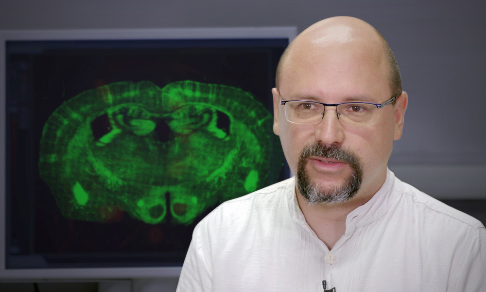
Alvaro Crevenna joined EMBL Rome in March 2019 as Head of the Microscopy Facility. He grew up in Mexico where he studied biomedical research and physics as an undergrad, and then followed a research career at several institutes in Europe. During his research he has gained considerable experience in using custom-built microscopy instruments to answer questions in cell biology and biophysics. He has always been keen on helping others to access technologies, so the transition to his current position has been a natural evolution in his career.
“Microscopy is an approach to understanding life, a window on the world, with a unique – perhaps biased – and sometimes colourful view,” says Alvaro. “Most of my academic career I’ve analysed time-lapse experiments, so science has been a movie. Maybe I just wanted to follow in the footsteps of my grandfather, who was a movie director and made more than 150 films.”
Building capacity by building a community
One of the main missions of the Microscopy Facility at EMBL Rome is to build a local network of microscopy users and collaborators. The facility provides users with training and support, including help with experimental design, image processing, and quantitative image analysis.
To serve a broader community, the facility is currently expanding its services by exporting know-how to partner institutes and engaging in technology transfer.
A collaboration with CrestOptics – a company specialised in microscopy technology – aims to develop super-resolution microscopy techniques for live cells and fixed tissue samples. EMBL Rome’s Microscopy Facility will serve as a platform for testing and providing insight on innovative imaging solutions, therefore accelerating the time to market.
Future omics directions
The long-term goal is to make the Microscopy Facility at EMBL Rome a centre of excellence for tissue imaging and image analysis. “To reach that aim, we need not only to foster early adoption of novel technology, but to participate directly in its development,” says Alvaro. For this reason, the facility is committed to developing microscopy-based omics approaches: platforms for multiplex in situ tissue profiling using proteomics (to detect tissue-specific proteins), transcriptomics (to identify genes that are expressed by specific cells in a tissue), and connectomics (to study how different areas of the brain are connected to each other). The goal is to carry out these different analyses on the same sample.
“A multi-plex tissue profiling platform established as a robust service for researchers would be unique in the world,” concludes Alvaro. “I’m really excited about the future discoveries that these techniques will bring forward.”
This article was first published on EMBL News