
EMBL Barcelona scientists have recapitulated for the first time in the laboratory how the cellular structures that give rise to our spinal column form sequentially
The spinal column is the central supporting structure of the skeleton in all vertebrates. Not only does it provide a place for muscles to attach, it also protects the spinal cord and nerve roots. Defects in its development are known to cause rare hereditary diseases. Researchers from the Ebisuya Group at EMBL Barcelona have now created a 3D in vitro model that mimics how the precursor structures that give rise to the spinal column form during human embryonic development.
The spinal column consists of 33 vertebrae, which form pairs of precursor structures called somites. Somites give rise to not only our vertebrae, but also our ribs and skeletal muscles. To ensure that these structures are formed correctly, somite development is tightly regulated, and each pair of somites arises at a particular sequential time point in development. This process is controlled by the segmentation clock, which is a group of genes that creates oscillatory waves, every wave giving rise to a new pair of somites.
For the first time, we have been able to create periodic pairs of human mature somites linked to the segmentation clock in the lab,” said Marina Sanaki-Matsumiya, first author of the study published in Nature Communications. Using this approach, the researchers developed a 3D in vitro model of human somite formation, also known as ‘somitogenesis’.
Time lapse of repetitive somite formation. Credit: Ebisuya Group/EMBL
Creating a robust somitogenesis process
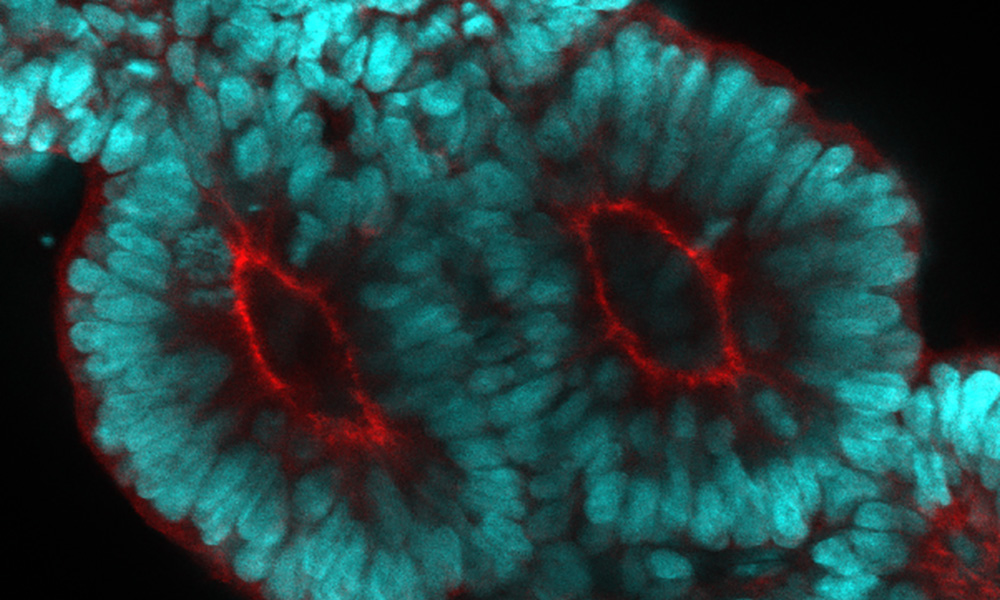
The team cultured human induced pluripotent stem cells (hiPSC) in the presence of a cocktail of signalling molecules that induce cell differentiation. Three days later, the cells started to elongate and create anterior (top) and posterior (bottom) axes. At that point, the scientists added Matrigel to the culture mix. Matrigel is what some scientists call the magic powder: a protein mixture that is critical to many developmental processes. This process eventually led to the formation of somitoids – in vitro equivalents of human somite precursor structures.
To test whether the segmentation clock regulates somitogenesis in these somitoids, the researchers monitored the expression patterns of HES7, the core gene involved in the process. They found clear evidence of oscillations, especially when somitogenesis was about to start. The somites that formed also had clear markers of epithelization – an important step in their maturation.
Somite size matters
The Ebisuya group studies how and why we humans are different from other species when it comes to embryonic development. One of the model systems they use to understand interspecies differences is the segmentation clock. In 2020, the group uncovered that the oscillation period of the human segmentation clock is longer than the mouse segmentation clock.
The current study also shows a link between the size of somites and the segmentation clock. “The somites that were generated had a constant size, independently of the number of cells used for the initial somitoid. The somite size did not increase even if the initial cell number did.” explained Sanaki-Matsumiya. “This suggests that the somites have a preferred species-specific size, which might be determined by local cell-cell interactions, the segmentation clock, or other mechanisms.”
To study this further, Miki Ebisuya and her group are now planning to grow somitoids of different species and compare them. The researchers are already working on several mammalian species, including rabbits, cattle, and rhinoceroses, setting up a ‘stem cell zoo’ in the lab.
“Our next project will focus on creating somitoids from different species, measure their cell proliferation and cell migration speed to establish what and how somitogenesis is different among species,” said Ebisuya.
Source article(s)
Periodic formation of epithelial somites from human pluripotent stem cells.
Sanaki-Matsumiya M., et al.
Nature communications 28th April
202210.1038/s41467-022-29967-1
This article was first published on EMBL News